Home
/ Anatomy Of Chest Bones / human thoracic skeleton bones max : A bone is a somatic structure that is comprised of calcified connective tissue.
Anatomy Of Chest Bones / human thoracic skeleton bones max : A bone is a somatic structure that is comprised of calcified connective tissue.
Anatomy Of Chest Bones / human thoracic skeleton bones max : A bone is a somatic structure that is comprised of calcified connective tissue.. They are always longer than they are wide the vertebrae are irregular bones. 12 photos of the anatomy bones chest. Bone basics and bone anatomy. It is made up of the wrist joint, the carpal bones, the metacarpal bones, and the phalanges. The mineral calcium phosphate hardens this framework, giving it.
Individuals may have more or fewer bones than the average (even accounting for developmental stage) owing to anatomical there are usually 25 bones in the chest but sometimes there can be additional cervical ribs in humans. Have you ever seen fossil remains of dinosaur and ancient human bones in textbooks, television, or in person at a museum? There also are bands of fibrous connective tissue—the ligaments and the tendons—in intimate relationship with the parts of the skeleton. Axial additionally, the scapula articulates with the chest wall to give the shoulder a greater net motion that. This webpage presents the anatomical structures found on wrist mri.
Human chest anatomy, illustration - Stock Image - F011 ... from media.sciencephoto.com Long bones are mostly located in the appendicular skeleton and include bones in the lower limbs (the tibia, fibula, femur, metatarsals, and phalanges) and bones in the upper limbs (the humerus, radius, ulna, metacarpals. Hand | definition, anatomy, bones, diagram, & facts. A collection of anatomy notes covering the key anatomy concepts that medical students need to learn. Where is the sternum found. Bone basics and bone anatomy. Bones of the chest and upper back (posterior view). An overview of the anatomy of the hand, including the bones of the hand, muscles, blood supply and nerve supply. The rib cage also anchors the bones of the head neck shoulders and arms to the trunk of the body.
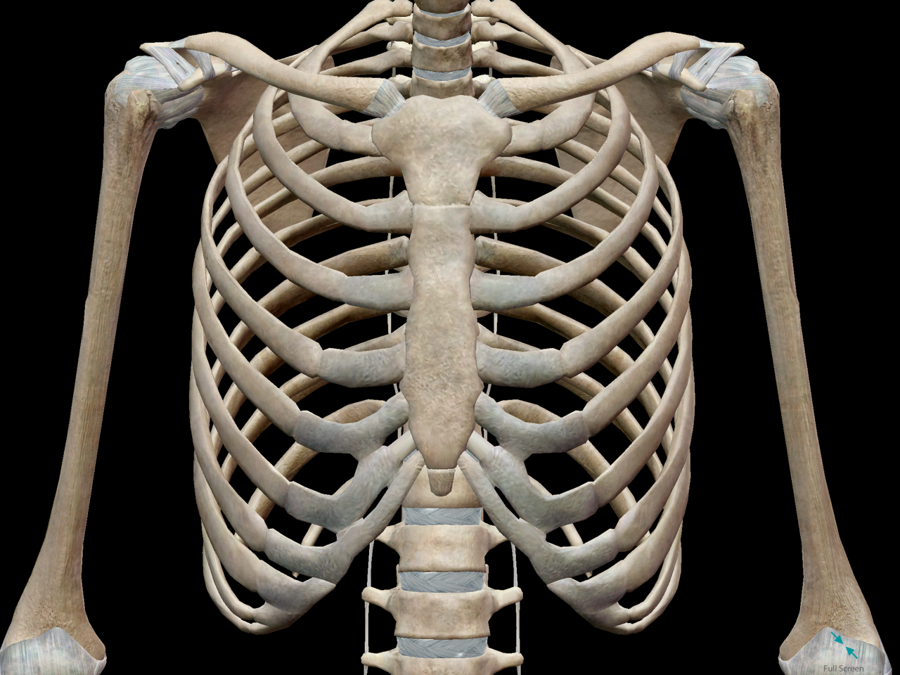
The ribs meet at an acute angle at the sternum, the costal cartilages thicken like beads at points of their transition to bones (rachitic beads).
Where is the sternum found. Part of a series of lists about. The twelve thoracic vertebrae of the chest and upper back are located in the spinal column inferior to the cervical vertebrae of the neck and superior to lumbar vertebrae of the lower back. Bones of the chest and upper back (posterior view). Ground substance and collagen fibers create a matrix that contains. Your rib cage, for example, acts like a shield around your chest to protect important organs inside such as your lungs and heart. Labeled chest radiographs teaching radiologic anatomy with a level of detail appropriate for medical students. When a patient flexes the neck forward, the prominent process is usually that of the 7th cervical. Long bones are categorised by their tubular shaft (diaphysis) with a rounded end (epiphysis) on each end. Bone basics and bone anatomy. The rib cage also anchors the bones of the head neck shoulders and arms to the trunk of the body. Bones are mostly made of the protein collagen , which forms a soft framework. It is comprised of many bones, formed by intramembranous ossification, which are joined together by sutures (fibrous joints).
Anatomists talk about both bone and bones. An overview of the anatomy of the hand, including the bones of the hand, muscles, blood supply and nerve supply. Atlas of anatomy of the human body: Computerized tomography 4 anatomy of lung segmental anatomy of lung lateral view on a normal lateral view the contours of the heart are visible and the ivc is seen entering the right atrium. The subchondral bone is not true cortical bone, in that it lacks some of the organization of cortical bone.
3D Skeletal System: Bones of the Thoracic Cage from www.visiblebody.com Bones have many shapes and sizes and are important to add structure to the body and protection to the vital structures. The mineral calcium phosphate hardens this framework, giving it. The skeleton is divided into 2 anatomic regions: This webpage presents the anatomical structures found on wrist mri. Long bones are categorised by their tubular shaft (diaphysis) with a rounded end (epiphysis) on each end. They are collectively known as the tarsus. Labeled chest radiographs teaching radiologic anatomy with a level of detail appropriate for medical students. The rib cage also anchors the bones of the head neck shoulders and arms to the trunk of the body.
This webpage presents the anatomical structures found on wrist mri.
Anatomists talk about both bone and bones. Individuals may have more or fewer bones than the average (even accounting for developmental stage) owing to anatomical there are usually 25 bones in the chest but sometimes there can be additional cervical ribs in humans. The mineral calcium phosphate hardens this framework, giving it. All of the anatomical and important histological facts about the bones, together with the clinical relations, are going to be desrcibed in this article. Bones have many shapes and sizes and are important to add structure to the body and protection to the vital structures. Related posts of anatomy bones chest. Your rib cage, for example, acts like a shield around your chest to protect important organs inside such as your lungs and heart. A bone is a somatic structure that is comprised of calcified connective tissue. Learn about each muscle, their locations & functional anatomy. The ribs meet at an acute angle at the sternum, the costal cartilages thicken like beads at points of their transition to bones (rachitic beads). The skeleton is divided into 2 anatomic regions: Anatomical illustrations of the lungs, chest, bronchi, trachea and thoracic lymph nodes. An overview of the anatomy of the hand, including the bones of the hand, muscles, blood supply and nerve supply.
Where is the sternum found. Talus calcaneus navicular cuboid lateral cuneiform intermediate cuneiform medial cune. These joints fuse together in adulthood, thus permitting brain growth during adolescence. Bones of the chest and upper back (posterior view). Individuals may have more or fewer bones than the average (even accounting for developmental stage) owing to anatomical there are usually 25 bones in the chest but sometimes there can be additional cervical ribs in humans.
GREAT website that shows you how to draw EVERYTHING ... from s-media-cache-ak0.pinimg.com Computerized tomography 4 anatomy of lung segmental anatomy of lung lateral view on a normal lateral view the contours of the heart are visible and the ivc is seen entering the right atrium. This webpage presents the anatomical structures found on wrist mri. Ground substance and collagen fibers create a matrix that contains. Individuals may have more or fewer bones than the average (even accounting for developmental stage) owing to anatomical there are usually 25 bones in the chest but sometimes there can be additional cervical ribs in humans. Long bones are categorised by their tubular shaft (diaphysis) with a rounded end (epiphysis) on each end. It, essentially, floats off of the back of the chest, as it is connected to the body primarily by muscle. Gross anatomy of axial skeleton. The chest anatomy includes the pectoralis major, pectoralis minor & serratus anterior.
Atlas of anatomy of the human body:
The ribs meet at an acute angle at the sternum, the costal cartilages thicken like beads at points of their transition to bones (rachitic beads). Have you ever seen fossil remains of dinosaur and ancient human bones in textbooks, television, or in person at a museum? All of the anatomical and important histological facts about the bones, together with the clinical relations, are going to be desrcibed in this article. There also are bands of fibrous connective tissue—the ligaments and the tendons—in intimate relationship with the parts of the skeleton. Your rib cage, for example, acts like a shield around your chest to protect important organs inside such as your lungs and heart. Anatomy of the chest wall. The scapula, or shoulder blade, is an approximately triangular shaped bone. Upper segment of sternum, flattened roughly triangular bone, o… the bony structure that forms the middle portion of the sternu… They are always longer than they are wide the vertebrae are irregular bones. These bones form by the thickening of a. Computerized tomography 4 anatomy of lung segmental anatomy of lung lateral view on a normal lateral view the contours of the heart are visible and the ivc is seen entering the right atrium. The bones of the chest and upper back combine to form the strong, protective rib cage around the vital thoracic organs such as the heart and lungs. They are collectively known as the tarsus.
The rib cage also anchors the bones of the head neck shoulders and arms to the trunk of the body anatomy of chest. It originates at your clavicle, ribs, and sternum, and inserts into the upper portion of your humerus (upper arm bone from elbow to shoulder.)

